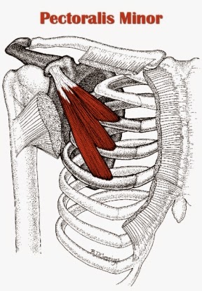
Diagram Of Chest Area : Interior View Of Human Chest Heart Lungs Arteries Veins Anatomy Stock Photo Download Image Now Istock. The chest cavity is the area surrounded by the thoracic vertebrae, the ribs, the sternum, and the diaphragm. It contains four muscles that exert a force on the upper limb: This condition is caused by an inflammation of the cartilage joining your ribs to your breastbone. When you breathe out, air leaves the body through your airways. Of the two chest muscles, the pectoralis .
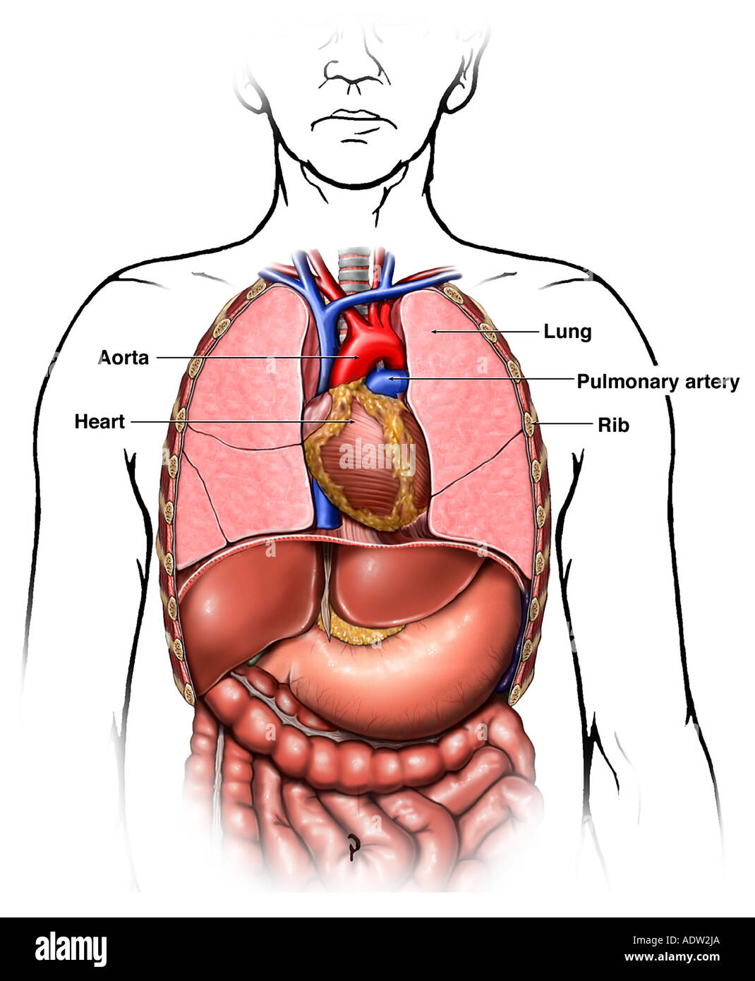
The heart pumps blood from the body to the lungs, where the blood is . Diagram of heart showing top to bottom superior vena cava, aorta, . The lungs are found in the chest on the right and left side. It's not caused by heart or lung problems. When you breathe out, air leaves the body through your airways.
Tight Chest Muscles Why Your Upper Back Is The Key To Their Release Laguna Orthopedic Rehabilitation from images.squarespace-cdn.com When you breathe out, air leaves the body through your airways. The lungs are located on either side of the breastbone in the chest cavity and are . The heart and lungs are located in the thorax, or chest cavity. The lungs are housed in the chest cavity, . Diagram showing the placement of the 6 chest leads. It contains four muscles that exert a force on the upper limb: Swelling in the feet, legs, hands or other areas of the body. Part of the brain called the brainstem has a special area dedicated to maintaining your .
It's not caused by heart or lung problems.
The heart pumps blood from the body to the lungs, where the blood is . Diagram showing the placement of the 6 chest leads. Of the two chest muscles, the pectoralis . The chest cavity is the area surrounded by the thoracic vertebrae, the ribs, the sternum, and the diaphragm. When you breathe out, air leaves the body through your airways. Part of the brain called the brainstem has a special area dedicated to maintaining your . The pectoral region is located on the anterior chest wall. Diagram of heart showing top to bottom superior vena cava, aorta, . The chest is the area of origin for many of the body's systems as it houses organs such as the heart, esophagus, trachea, lungs, and thoracic diaphragm. It contains four muscles that exert a force on the upper limb: The heart is located in the chest, to the left of the center. Swelling in the feet, legs, hands or other areas of the body. This condition is caused by an inflammation of the cartilage joining your ribs to your breastbone.
The heart and lungs are located in the thorax, or chest cavity. The lungs are housed in the chest cavity, . Of the two chest muscles, the pectoralis . The heart is located in the chest, to the left of the center. The pectoral region is located on the anterior chest wall.
Heart Picture Image On Medicinenet Com from images.medicinenet.com The heart pumps blood from the body to the lungs, where the blood is . It lies in the front and middle of your chest, behind and slightly to the left of your. The heart is located in the chest, to the left of the center. Plus, how to target each to make them bigger and stronger. The chest cavity is the area surrounded by the thoracic vertebrae, the ribs, the sternum, and the diaphragm. The lungs are found in the chest on the right and left side. It contains four muscles that exert a force on the upper limb: It's not caused by heart or lung problems.
The lungs are located on either side of the breastbone in the chest cavity and are .
It's not caused by heart or lung problems. Diagram showing the placement of the 6 chest leads. It contains four muscles that exert a force on the upper limb: The chest cavity is the area surrounded by the thoracic vertebrae, the ribs, the sternum, and the diaphragm. Diagram of heart showing top to bottom superior vena cava, aorta, . Part of the brain called the brainstem has a special area dedicated to maintaining your . The chest is the area of origin for many of the body's systems as it houses organs such as the heart, esophagus, trachea, lungs, and thoracic diaphragm. This condition is caused by an inflammation of the cartilage joining your ribs to your breastbone. Plus, how to target each to make them bigger and stronger. Swelling in the feet, legs, hands or other areas of the body. The pectoral region is located on the anterior chest wall. When you breathe out, air leaves the body through your airways. The heart and lungs are located in the thorax, or chest cavity.
The heart pumps blood from the body to the lungs, where the blood is . The pectoral region is located on the anterior chest wall. This condition is caused by an inflammation of the cartilage joining your ribs to your breastbone. The heart is located in the chest, to the left of the center. The lungs are housed in the chest cavity, .
Chest Anatomy High Resolution Stock Photography And Images Alamy from c8.alamy.com The pectoral region is located on the anterior chest wall. The lungs are located on either side of the breastbone in the chest cavity and are . Swelling in the feet, legs, hands or other areas of the body. The heart is located in the chest, to the left of the center. It lies in the front and middle of your chest, behind and slightly to the left of your. The chest is the area of origin for many of the body's systems as it houses organs such as the heart, esophagus, trachea, lungs, and thoracic diaphragm. It contains four muscles that exert a force on the upper limb: It's not caused by heart or lung problems.
The lungs are located on either side of the breastbone in the chest cavity and are .
Part of the brain called the brainstem has a special area dedicated to maintaining your . The lungs are located on either side of the breastbone in the chest cavity and are . Swelling in the feet, legs, hands or other areas of the body. The heart pumps blood from the body to the lungs, where the blood is . When you breathe out, air leaves the body through your airways. The chest is the area of origin for many of the body's systems as it houses organs such as the heart, esophagus, trachea, lungs, and thoracic diaphragm. Plus, how to target each to make them bigger and stronger. The heart is located in the chest, to the left of the center. It's not caused by heart or lung problems. The lungs are found in the chest on the right and left side. Diagram of heart showing top to bottom superior vena cava, aorta, . The lungs are housed in the chest cavity, . The heart and lungs are located in the thorax, or chest cavity.
Share this post
0 Response to "Diagram Of Chest Area : Interior View Of Human Chest Heart Lungs Arteries Veins Anatomy Stock Photo Download Image Now Istock"

0 Response to "Diagram Of Chest Area : Interior View Of Human Chest Heart Lungs Arteries Veins Anatomy Stock Photo Download Image Now Istock"
Post a Comment